|
Always get a Pre Purchase ExamThe equine pre purchase exam will inform the buyer of existing or potential problems that could render the horse unsuitable for its intended use. This article explains what a veterinarian examines when conducting a pre purchase exam. Unlike other consumer products that one might purchase, horses rarely come with a guarantee. Therefore, the buyer must make the best possible attempt to select an animal that is suitable and capable of performing a given function. The final determination, of course, is the price. The combination of function capability, suitability and price enter into the ultimate decision of whether a prospective buyer should purchase a given animal.
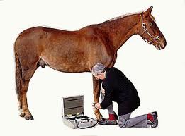
The purchase examination or pre purchase examination, done by a qualified veterinarian, is perhaps one of the best ways to help the prospective buyer determine serviceability. It is the veterinarian’s responsibility to look for physical and medical evidence to determine whether the horse will withstand a particular use for the prospective buyer. The term “pre purchase examination” implies a complete clinical examination that includes consideration of the pertinent medical history and environmental factors. All other terms that have been associated with this type of examination should be abandoned because they are either incomplete or cause confusion about the type of examination. The role of the veterinarian is to find medical problems on the date of the pre purchase exam and evaluate conformation defects in light of problems that they may cause, as well as evaluating overall health. Even though the procedure is applicable to all classes of horses, the veterinarian involved should have a thorough knowledge of the class and use of the horse to be examined.
It is inadvisable to examine and determine the potential of an animal that is not on a regulated work schedule. The immature animal that is not in training must be given extended paddock exercise in order to completely evaluate the musculoskeletal and cardiopulmonary systems. It is not the domain of the examining veterinarian to determine the presence of undesirable traits or vices. The disclosure of this information is dependent upon the integrity of the seller.
Oral and Written Reports An oral or written report is permissible, depending on the desires of both the veterinarian and the prospective buyer. However, it is imperative that the animal examined is completely identified for the pre purchase exam. In addition, a record of all clinical findings, including radiographs (X-rays) if taken, must be maintained in the permanent office files of the examining veterinarian.
It is important that each veterinarian develop a standardized basic procedure in the pre purchase exam of the horse for purchase. More detailed diagnostic techniques are performed in the event of suspicious disease problems or client request. In these cases, consultation with a specialist may be required.
Before the pre purchase exam is begun, any history of preventive medicine programs and previous medical problems is obtained. The horse’s markings and other means of identification are noted. Ideally, the horse has been worked by the prospective buyer before the pre purchase exam so it is evident that horse and rider are suited for each other. Findings should be recorded in the order noted so the entire examination is not trusted to memory.
During the pre purchase exam the horse is viewed in its stall for evidence of abnormal stance or posture and temperament. The horse’s eyes are then examined with a direct ophthalmoscope. An indirect, aspheric viewing lens with a halogen-illuminated light source is excellent for a panoramic view of the ocular fundus (rear portion of the eye). Ophthalmic abnormalities are interpreted in terms of the horse’s intended use. The transilluminator of the ophthalmoscope is used to examine the nasolacrimal puncta and external ear canals. The mouth is rinsed and examined, manually and with the transilluminator, for dental disease, malocclusion or other abnormalities of the oral cavity.
With the horse still in the stall, the rectal temperature is measured and the respiratory rate and pattern noted. The heart is monitored with a stethoscope from both sides while the transverse facial or facial artery are palpated for irregularities of rate or rhythm. Occluding the horse’s nostrils for at least 30 seconds or using a large plastic bag over the nostrils often accentuates respiratory problems as indicated by subsequent coughing or dyspnea. This technique also causes the horse to breathe deeply for thorough monitoring of the lungs. The trachea and larynx are monitored carefully and the nasal sinuses tapped to diagnose the condition of the underlying parts.
The horse is then taken to a well-lit, level, firm surface and made to stand with its weight equally distributed on all four legs. During this part of the pre purchase exam the horse’s conformation, balance and symmetry from all views, are carefully evaluated, with faults predisposing to lameness or gait abnormalities noted.
The Head In the next part of the pre purchase exam, the head is observed for symmetry, carriage and coordination of movement, and any cranial nerve defects noted. The mandibular lymph nodes, parotid glands and guttural pouch areas are palpated. The larynx is palpated for atrophy of the cricoarytenoideus dorsalis muscle or signs of previous surgery. The laryngeal cartilages often can be compressed, causing inspiratory noise in horses with laryngeal hemiplegia (roaring). The ventral neck muscles are palpated and the patency of both jugular veins determined. The cervical vertebrae neck bones are palpated, beginning at the poll. Symmetry of the wings of the atlas (first cervical vertebra) and the horse’s response to cervical vertebral palpation and manipulation are noted.
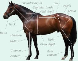
The Body Examination of the body begins on the left side with palpation of the spine of the scapula, point of the shoulder, and muscles of the shoulder and chest. Working posteriorly on the topline, the withers, dorsal spinous processes of the vertebra, tuber sacrale and point of the hip are palpated. The muscles of the back, loin and croup are palpated, and evidence of muscle guarding noted. During the pre purchase exam the girth region and ventral abdomen are examined for midline dermatitis, absence of the umbilicus or signs of previous abdominal surgery. Several breeds of horses will not allow the exhibition of horses with altered tail function. An electromyogram may be necessary to prove normal tail function. Normal tail tone and function should be determined by manipulation and careful observation when the horse is worked. Evidence of coccygeal muscle atrophy and surgical or medical alteration of normal tail function should be noted; the gluteal and caudal thigh muscles are palpated and compared for symmetry. Unilateral muscle atrophy may indicate chronic rear-leg lameness. The external genitalia are also examined at this time. Examination of the body is then repeated on the right side.
The Legs Next, each limb is inspected, beginning with palpation of the structures of the left forelimb. The digital pulse (pulse at the posterior fetlock) is taken before the sulci and the frog are cleaned and the sole pared, if necessary. The comparative size, balance, wear and quality of each hoof are noted. A flat sole, atrophied frog, divergent growth rings, sheared heels, improper hoof-pastern angle and dished cranial hoof wall may indicate previous or existing foot disease. The type of shoe, presence of pads and shoe wear may also provide useful information. The pliability of the collateral cartilages of the coffin bone and presence of coffin joint excess fluid are determined during the pre purchase exam. The posterior pastern region is examined for neurectomy scars, painful neuromas, digital sheath effusion, thickening of the distal sesamoidean ligaments and ringbone. Firm pressure applied to either side of the extensor tendon on the face of the fetlock may reveal villonodular lesions (proliferative nodules), extensor process fractures, or osselets (bony protrusions). Fetlock joint effusion, windpuffs and annular ligament constriction are conditions commonly encountered.
While the horse's leg is flexed at the knee, each sesamoid bone and its branch of the suspensory ligament is palpated for thickening, enlargement or pain. Then the suspensory ligament is palpated to its origin and the flexor tendons palpated for thickening, pain and loss of separation along their lengths. During palpation of the suspensory ligament, abnormal enlargements of the splint bones are often encountered. With the limb still flexed, each splint bone should be palpated from its distal end to its proximal attachment. Pain on palpation of splint bone enlargements and degree of impingement on the suspensory ligament should be carefully considered. With the leg flexed, the carpal bones are palpated for thickening or tears of the fibrous joint capsule and synovial effusion. The accessory carpal bone and carpal canal are also palpated. Proceeding from the carpus, the muscles of the forearm, distal radius, chestnut and olecranon are inspected. The range of motion and response to forced flexion or rotation of the digit, pastern, fetlock and carpus are determined after palpation of the forelimb. The shoulder and elbow usually are not manipulated. Decreased range of motion and abnormal response to manipulation are noted and reevaluated when the horse is examined in motion.
Examination of the hindlimb below the hock is similar to that of the forelimb. At the hock, effusion of the tarsal sheath, tibiotarsal joint and tendon sheaths over the face of the hock are evaluated. The bony structures of the hock, point of the hock and plantar ligament are palpated. The surgical sites for cunean tenotomy and lateral digital extensor tenotomy are examined. Proceeding to the stifle joint, the tibial crest, patellar ligaments, patella and lateral trochlea of the femur are palpated. Horses with chronic upward fixation of the patella often resent palpation of the lateral trochlea of the femur as well as attempts to fix the patella. By this time in the pre purchase exam, the horse’s temperament should be evident. Most horses permit the examiner to stand behind them and palpate both stifle joints simultaneously, allowing comparison of synovial effusion and joint capsule thickening. The medial collateral ligament of the stifle should also be palpated. After the structures of the hindlimb are examined, the digit, pastern, fetlock, hock and stifle are manipulated for range of motion and pain.
When the body and legs have been examined, hoof testers can be applied to the feet. Occasionally, a seller may implicate the hoof testers as causing lameness. For this reason, examination with hoof testers is done after the horse has been observed at work.
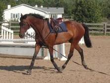
Timed flexion or other manipulative tests are performed after work on the longe line, with emphasis on areas with suspected problems. The response to forced flexion, rotation or digital pressure varies greatly between individuals and must be considered carefully, along with the history and clinical findings. The horse is then watched at work under saddle. After a warm-up period, the horse is observed at a walk, trot and canter. Trotting in a tight figure-8 pattern frequently reveals subtle lameness. Lead changes and length of stride can be evaluated at a canter.
Though it is the trainer’s job to determine whether the horse’s disposition and athletic ability are suited to the rider and intended use, it is very informative to watch the horse perform as intended. Tail-wringing, ear-pinning, unwillingness to work in one direction, cross-cantering, failure to pick up leads or other inappropriate behavior often indicate problems. After the horse is worked hard, the larynx and heart are auscultated. Heart and respiratory rates are determined during recovery from exercise.
Other Procedures Once the physical part of the pre purchase exam is completed, other diagnostic procedures may be done. Radiography is probably the most common ancillary procedure performed. Radiographs are not routinely obtained in the absence of clinical findings, unless requested by the buyer. Other special diagnostic procedures performed include endoscopic examination of the upper respiratory system, fecal exam for internal parasite ova, rectal examination, uterine biopsy and breeding soundness examination. Blood is drawn for a Coggins’ test for equine infectious anemia, if not current. Blood chemistry analyses, a CBC, urinalysis, drug tests and an electrocardiogram and cardiac ultrasound are other tests that are mentioned for the sake of completeness. However, certain situations may warrant the use of these tests.
Prospective buyers should also be made aware of the potential for genetic problems in certain breeds/sire lines of horses. An example is Hyperkalemic Periodic Paralysis (HyPP). HyPP is an inherited defect seen in descendants of a particular Quarterhorse sire characterized by intermittent episodes of muscular fasiculations, weakness, mytonia and/or involuntary recumbency. During these episodes the serum potassium is often elevated but between occurrences the potassium concentration is normal. For affected horses, this disease is a lifelong problem. This disease is an autosomal dominant trait. Owners and breeders with affected horses should inform prospective buyers of the potential for this disease to occur. There are four methods for confirming HyPP—hyperkalemia during an episode, a potassium chloride challenge, electromyography and testing for a horse-specific sodium channel genetic marker.
After the entire pre purchase exam is complete, the veterinarian may offer a prognosis to the buyer. In considering the prognosis for a performance horse, one must consider the horse’s conformation, temperament, intended use, past history, clinical problems and results of ancillary diagnostic procedures.
The purpose of the pre purchase exam is not to pass or fail the horse but to inform the buyer of existing or potential problems that could render the horse unsuitable for its intended use. Certain warranties or agreements between the buyer and seller such as the length of serviceability, performance ability and avoidance of drugs before the examination -- are not the responsibility of the veterinarian. The relatively small investment at the time of purchase for a good quality examination can help prevent a much larger investment in treatment or loss of performance later on. Such an exam is certainly a good choice for an informed purchase.
SOURCE - G. Anderson
|