|
Common Lameness in Horses
This article discusses three types of common lameness in horses: proximal suspensory desmitis, tarsitis and patellar fixation.
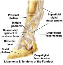
Proximal Suspensory Desmitis The suspensory ligament is an important supporting structure which lies on the flexor surface of the cannon region of the fore and hindlimb. It originates from the palmar/plantar surface of the proximal cannon bone and inserts on the proximal sesamoid bones. Although injury can occur to any part of the suspensory ligament, desmitis of the proximal portion at its attachment to the cannon bone is particularly difficult to manage.
Onset of clinical signs may be insidious or sudden and may manifest as mild to severe lameness in Horses. The condition may be predisposed by straight hock conformation. The diagnosis is confirmed by diagnostic nerve blocks and ultrasound.
Most horses are treated by stall rest with controlled exercise consisting of gradual increase in work load and ultrasound re-evaluations every 90 days. However, this approach alone seldom results in return to soundness. Extracorporeal shockwave therapy has become popular and reports indicate about 50-percent success rate. Injection of the lesion with bone marrow harvested from the horse’s sternum has also been reported, with variable results.
Recently, a combination of splitting the suspensory origin and performing a fasciotomy under ultrasonic guidance has been proposed. The treatment is based on the theory that the enlarged suspensory ligament is trapped under the overlying fascial planes and that the surgery will relieve the compression of these structures (“compartment syndrome”). A similar procedure includes performance of a neurectomy of the deep branch of the lateral nerve as well as a fasciotomy. Initial reports of success for these two procedures have been high but will need the test of time. Regardless of the treatment, the prognosis for these cases is guarded, particularly in the hindlimb.
The diagnosis is largely based on the Churchill hock test, which involves putting digital pressure over the first and second tarsal bones and the head of the inside splint bone in the non-weight bearing limb. A positive response is indicated by limb abduction (not flexion).
Intra-articular anesthesia can also be used to confirm the diagnosis. Radiographs correlate very poorly to clinical signs and are rarely, if ever, helpful in making the diagnosis of lameness in horses. Intra-articular injections of corticosteroids or hyaluronan are usually effective in alleviating the signs. NSAIDs, such as phenylbutazone, are often helpful, as are anti-arthritic drugs such as Adequan or Legends and nutraceuticals containing glucosamine and chondroitin sulfate. Attention should be paid to shoeing. For example, it is often helpful to shorten the toe to aid breakover. It is important that any underlying primary lameness in horses be identified and treated concurrently. More persistent cases of tarsitis with significant osteoarthritis may be candidates for cunean tenectomy or surgical arthrodesis (fusion) of the affected joints. Prognosis is generally good for most cases and even persistent cases can often be managed medically.
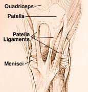
Patellar fixation There are two manifestations of this problem. First is the classical form, in which the horse has its stifle locked in extension and drags its toe as a result. This is more common in miniatures, ponies and horses that are confined. A far more common form has been referred to as delayed release of the patella. It is an intermittent form in which the patella catches occasionally, followed by a “popping” of the stifle or mildly exaggerated flexion of the stifle or hock. This form accounts for about 95-percent of stifle lameness in horses.
As a normal part of stifle anatomy and function, the medial patellar ligament attaches to a fibrocartilage extension of the patella which catches in the notch at the top of the medial trochlea. Relaxation of the extensor muscles allows slight flexion of the stifle, which locks the patella in place. When the horse wants to move forward, the biceps femoris muscle contracts, rotates the patella laterally, and disengages the medial patellar ligament. Then the flexor muscles flex the joint. When this mechanism fails, the patellar release is delayed momentarily, producing the clinical signs.
This lameness in horses is extremely common and is seen in all types of horses. It is almost always bilateral, although the lameness is usually noticed in only one limb. Radiographs and diagnostic joint blocks are not helpful in making the diagnosis. The patellar fixation test is performed, in which caudal or backward pressure is applied to the patella and the horse is taken ahead one step. A catching of the patella indicates a positive test.
Patellar fixation is effectively treated with injections of 2-percent iodine in oil into the middle and medial patellar ligaments. The horse is returned to work with plenty of trotting. Most horses require only one treatment, but occasionally repeated treatments are necessary. The prognosis is good.
Surgical treatment, which involves medial patellar desmotomy, should be reserved only for very severe or resistant cases. It is essential that the horse be given two to five months rest after the surgery. A common sequel to desmotomy is fragmentation of the patella, which can result in severe lameness that may require arthroscopic surgery.
SOURCE - M. P. Brown
Return from Lameness in Horses to Horse Riding Connection
|
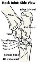 Tarsitis (distal hock pain) The hock joint (tarsus) actually consists of four joints from proximal to distal: tibiotarsal joint (tarsocrural), proximal intertarsal joint, distal intertarsal joint and tarsometatarsal joint. The latter two most distal joints are most commonly involved in lameness in horses. Distal hock pain is seen in all types of horses. Onset may be sudden or gradual, with varying degrees of lameness. It may be unilateral or bilateral and is often secondary to other lameness problems, such as sore front feet. It may be in the form of synovitis or osteoarthritis.